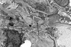
Electron microscopy of a biopsy specimen in a patient with IgA nephropathy. Electron dense deposits can be identified in the mesangium (black arrows), which on immunofluorescence would have predominant or co-dominant IgA staining. Note that although mesangial IgA deposits are classic in IgA nephropathy and IgA vasculitis (former Henoch-Schonlein purpura…
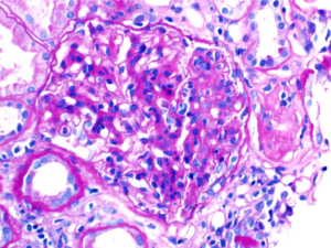
A patient with IgA nephropathy and associated segmental glomerulosclerosis. Images courtesy of Patrick Walker, MD.
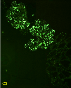
Immunofluorescent staining pattern in a patient with C3 glomerulopathy. There is strong C3 staining in the capillary loops and mesangium. Immunoglobulin staining, such as IgG, is typically absent or at a lower intensity by at least two orders of magnitude than C3. Images courtesy of Patrick Walker, MD.