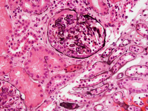
IgA nephropathy
IgA nephropathy with crescents. Note the fibrocellular crescent present in this glomerulus extending from the 3 o’clock the 12 o’clock position. Though difficult to appreciate in this image, there is…
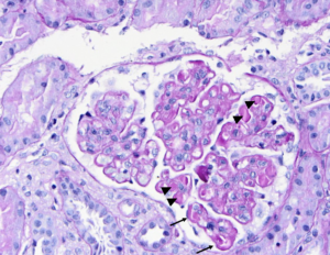
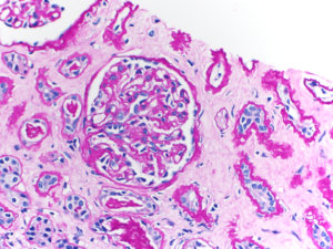
SLE nephritis
PAS stain of a glomerulus in a patient with SLE nephritis. There is endocapillary and mesangial proliferation, as evidenced by thickened, occlusive capillary loops and increased mesangial cellularity. Note the…
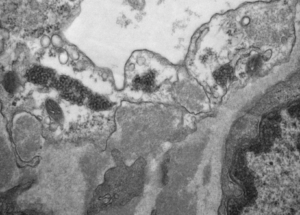
Tubuloreticular inclusions in SLE nephritis
Tubuloreticular inclusions in a patient with diffuse proliferative SLE nephritis (SLE class IV). These subcellular structures (dark circular clusters) on transmission electron microscopy are localized to the cytoplasm of endothelial…
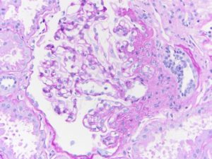
Focal and segmental glomerulosclerosis
Segmental obliteration of the glomerular capillary lumen in a patient with FSGS. Note the sclerotic portion of the glomerular tuft is adherent to Bowman’s capsule. There is proximal tubular hypertrophy,…
Transplant glomerulopathy
The glomerular capillary bed is thickened, with the presence of double contours reflecting circumferential mesangial expansion into the capillary wall (mesangial interposition). In this biopsy core of chronic allograft injury…
- « Previous
- 1
- 2
- 3