A normal glomerulus (left) and hypertrophied glomerulus (glomerulomegaly, right). Glomerulomegaly is an adaptive response to decreased nephron number (e.g. prematurity) and/or increased demand (e.g. obesity). Patients with glomerulomegaly may have sub-nephrotic or nephrotic-range proteinuria, but other features of nephrotic syndrome are rare. Images courtesy of Patrick Walker, MD.
Read More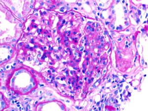
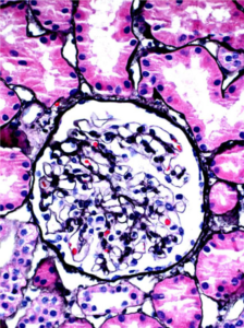
A patient with IgA nephropathy and associated segmental glomerulosclerosis. Images courtesy of Patrick Walker, MD.
Read More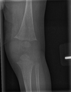
Early radiographic changes on left knee X-ray in an infant with hypophosphatemic rickets. There is decreased mineralization of the long bones and splaying, or widening, at the distal femur and proximal tibia, where the metaphyses are located.
Read More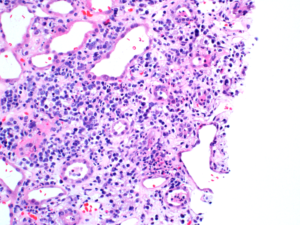
A mixed inflammatory cell infiltrate in a child with acute interstitial nephritis. Image courtesy of Patrick Walker, MD.
Read More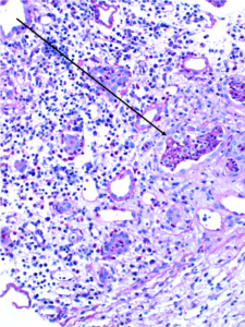
WBC casts (black arrows) in a biopsy specimen of a patient with acute interstitial nephritis. Images courtesy of Patrick Walker, MD.
Read More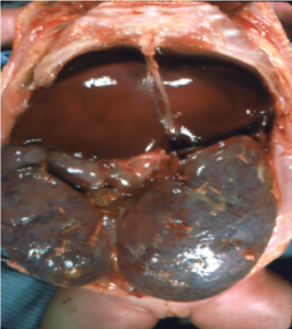
Gross pathology and low-power light microscopy of kidney tissue in a neonate with ARPKD. The kidneys are enlarged but maintain their reniform shape, and are full of microscopic cysts derived from dilated distal tubules and cortical collecting ducts. Images courtesy of Patrick Walker, MD.
Read More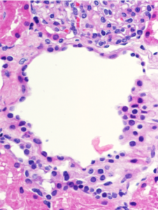
Microscopic renal cysts. Note the flattened to cuboidal epithelium lining the cysts. Images courtesy of Patrick Walker, MD.
Read More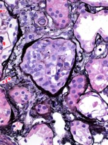
A segmental (left, black arrow) and circumferential crescent (right) in a patient with IgA nephropathy. Images courtesy of Patrick Walker, MD.
Read More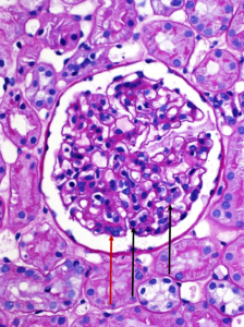
A normal appearing glomerulus (left) compared to a glomerulus with endocapillary hypercellularity (right). Note the hypercellular capillary loop (red arrow) compared to the normal capillary lumens (black arrows). This histologic feature can be seen in several glomerular disorders, including IgA nephropathy, post-infectious glomerulonephritis, lupus nephritis, and C3 glomerulopathy. Images courtesy…
Read More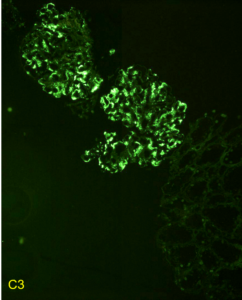
Immunofluorescent staining pattern in a patient with C3 glomerulopathy. There is strong C3 staining in the capillary loops and mesangium. Immunoglobulin staining, such as IgG, is typically absent or at a lower intensity by at least two orders of magnitude than C3. Images courtesy of Patrick Walker, MD.
Read More