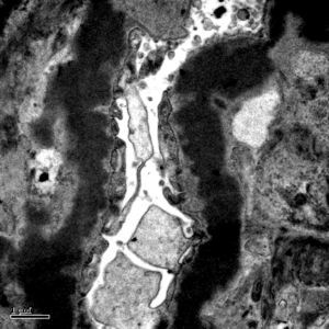
Dense deposit disease on electron microscopy in a patient with C3 glomerulopathy. This lesion results from intramembranous transformation of the glomerular basement membrane by sausage-like, “osmiophilic” dense material. Images courtesy of Patrick Walker, MD.
Read More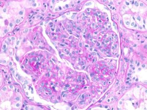
Membranoproliferative pattern of glomerular injury in a patient with C3 glomerulopathy. This pattern of injury typically has endocapillary proliferation, diffuse capillary wall thickening, increased mesangial matrix, and mesangial proliferation visible on light microscopy, producing a lobular appearance of the glomerular tuft. Images courtesy of Patrick Walker, MD.
Read More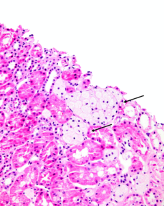
Clusters of interstitial foam cells (arrows) in a kidney biopsy. These are commonly found in biopsy specimens of patients with Alport syndrome, FSGS, IgA nephropathy, and other proteinuric kidney diseases. Image courtesy of Patrick Walker, MD.
Read More
For many in our community, the events of the last week have caused great consternation and triggered feelings of intense disappointment, frustration, and anger. For others, what has transpired has caused frank fear for the safety of colleagues, friends, family, and loved ones as well as concern for those whom…
Read More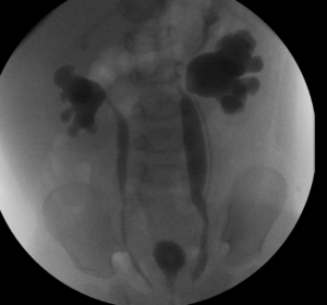
This voiding cystourethrogram shows bilateral vesicoureteral reflux grade 5. Also, on left side, two separate ureters can be seen.
Read More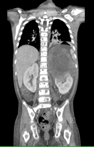
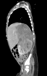
This image shows a large neuroblastoma mass at upper pole of left kidney, distorting its architecture
Read More
We are very sad to announce the death of Professor Michel Broyer on March 10, 2020 at the age of 86, following a COVID19 infection. Early during his residency in Professor Pierre Royer’s unit at the Necker-Enfants Malades Hospital in the early 1960’s, Michel Broyer was fascinated by the recent…
Read More