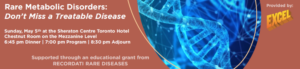
Join us for an enlightening CME Satellite Symposium focusing on rare metabolic disorders. This educational event aims to raise awareness among pediatricians about the importance of newborn screening, diagnosis, and management of these often-overlooked disorders. Date: Sunday, May 05, 2024 Location: Pediatric Academic Societies Annual Meeting, Metro Toronto Convention Center,…
Read More
We are excited to announce a new feature on the ASPN Go app! You can now post directly on the newsfeed to share updates, insights, and engage with fellow members. How to Post on the Newsfeed: 1. Open the ASPN Go app. 2. Navigate to the ‘Newsfeed’ section. 3. Tap…
Read More

Thank you for your interest in accessing our exclusive content. Please note that this section is reserved for ASPN members only. If you're already a member, please log in using your credentials to unlock access using the button below.
If you're not yet a member or if your membership has expired, don't worry! You can easily sign up or renew your membership to gain immediate access to all our valuable resources and benefits.
To sign up or renew your membership, simply click here "Sign Up" or "Renew Membership", and follow the instructions.

Remembering Patrick “Pat” Brophy, a transformational leader, whose focus was always on the children. Patrick (“Pat”) D. Brophy, MD, MHCDS, beloved husband, father, brother, friend, and leader, left us all too soon on October 6, 2023, after a long battle with lymphoma. While the facts of his life can be…
Read More
Thank you for your interest in accessing our exclusive content. Please note that this section is reserved for ASPN members only. If you're already a member, please log in using your credentials to unlock access using the button below.
If you're not yet a member or if your membership has expired, don't worry! You can easily sign up or renew your membership to gain immediate access to all our valuable resources and benefits.
To sign up or renew your membership, simply click here "Sign Up" or "Renew Membership", and follow the instructions.