
Dr. Ronald Jay Portman passed away peacefully on May 30, 2024 surrounded by his loving family. He is survived by the love of his life for 50+ years, Joan (Joni) Portman. Together they had three children, Wendi Portman Fried, Shayna Portman, and Zack Portman whose spouses, his sons and daughter…
Read More

Thank you for your interest in accessing our exclusive content. Please note that this section is reserved for ASPN members only. If you're already a member, please log in using your credentials to unlock access using the button below.
If you're not yet a member or if your membership has expired, don't worry! You can easily sign up or renew your membership to gain immediate access to all our valuable resources and benefits.
To sign up or renew your membership, simply click here "Sign Up" or "Renew Membership", and follow the instructions.
The Pediatric Centers of Excellence in Nephrology proudly announce: The Call for Pilot and Feasibility Proposals The Pediatric Centers of Excellence in Nephrology (PCENs) at the University of Virginia, Children’s Hospital of Philadelphia, and Washington University in St. Louis are issuing a joint RFA for Pilot and Feasibility (P&F) projects. The PCEN program is…
Read More
Thank you for your interest in accessing our exclusive content. Please note that this section is reserved for ASPN members only. If you're already a member, please log in using your credentials to unlock access using the button below.
If you're not yet a member or if your membership has expired, don't worry! You can easily sign up or renew your membership to gain immediate access to all our valuable resources and benefits.
To sign up or renew your membership, simply click here "Sign Up" or "Renew Membership", and follow the instructions.
Join us on this final episode of the The Sediment to discuss the 2024 Pediatric Academic Societies, American Society of Pediatric Nephrology (ASPN) conference sessions on urinary tract infections, vaccinations in nephrotic, CKD and transplant patients, hypertension, and neonatal nephrology with their respective moderators. Hot-off-the-press JAMA Peds article “Long-Term Cardiovascular Outcomes in Children…
Read More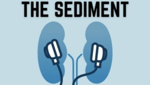
Join us on this episode of The Sediment to discuss the 2024 Pediatric Academic Societies, American Society of Pediatric Nephrology (ASPN) conference sessions on kidney transplantation in special patient populations, big ideas and small numbers research, chronic dialysis and critical care nephrology with their respective moderators. St. Jude Childhood Cancer…
Read MoreJoin us on this episode of The Sediment to discuss the 2024 Pediatric Academic Societies, American Society of Pediatric Nephrology (ASPN) conference sessions on kidney transplantation in special patient populations, big ideas and small numbers research, chronic dialysis and critical care nephrology with their respective moderators. St. Jude Childhood Cancer…
Read More