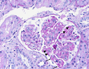
PAS stain of a glomerulus in a patient with SLE nephritis. There is endocapillary and mesangial proliferation, as evidenced by thickened, occlusive capillary loops and increased mesangial cellularity. Note the “wire loop” lesions (arrows) and hyaline thrombi present (arrowheads). Image courtesy of Joseph Gaut, MD, PhD.
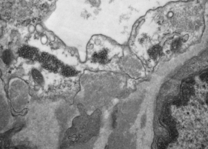
Tubuloreticular inclusions in a patient with diffuse proliferative SLE nephritis (SLE class IV). These subcellular structures (dark circular clusters) on transmission electron microscopy are localized to the cytoplasm of endothelial cells, and thought to be formed in high interferon states. These are classic for SLE nephritis, but can be seen…
- « Previous
- 1
- 2
- 3