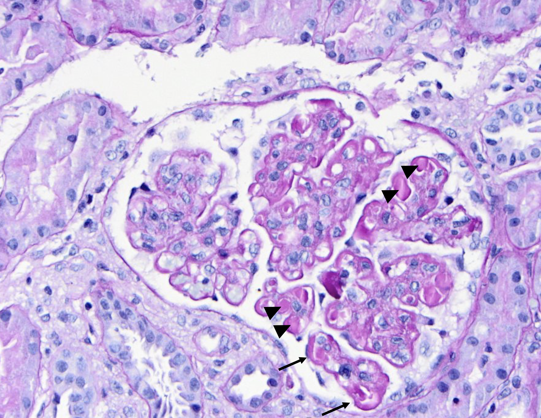
PAS stain of a glomerulus in a patient with SLE nephritis. There is endocapillary and mesangial proliferation, as evidenced by thickened, occlusive capillary loops and increased mesangial cellularity. Note the “wire loop” lesions (arrows) and hyaline thrombi present (arrowheads). Image courtesy of Joseph Gaut, MD, PhD.

Posted: February 27, 2024
Posted in Glomerular Diseases