Calyceal Diverticulum on MRU


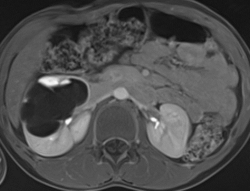
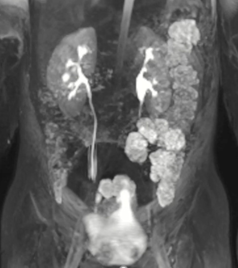
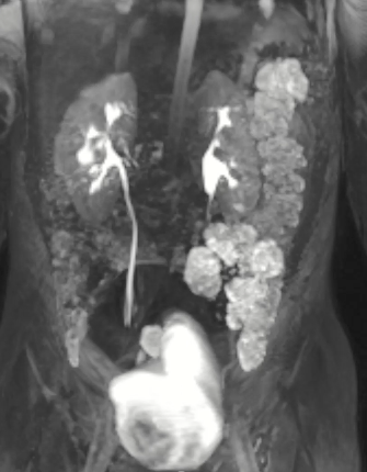
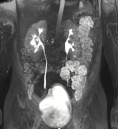
Right anterior calyceal diverticulum on functional MR Urography. Top left side shows a T2 weighted image (no contrast) where fluid is bright, top right image is 10 min post-contrast on axial plane, and bottom images are a coronal views showing filling of diverticulum on dynamic phase. Images courtesy of Hansel Otero, MD.

NKC Educational Symposium: Fetal Interventions for CAKUT from a Multidisciplinary Perspective
October 22 @ 3:00 pm - 5:00 pm EDT
ASPN Policy Update
October 23 @ 12:00 pm - 1:00 pm EDT
Board Review Course 2025
October 29 - October 30
ASPN Workforce Committee Meeting
October 29 @ 1:00 pm - 2:00 pm EDT
ASPN Advanced Practice Providers SIG Quarterly Meeting
October 29 @ 4:00 pm - 5:00 pm EDT
Posted in