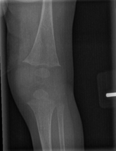
Early radiographic changes on left knee X-ray in an infant with hypophosphatemic rickets. There is decreased mineralization of the long bones and splaying, or widening, at the distal femur and proximal tibia, where the metaphyses are located.
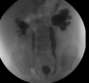
This voiding cystourethrogram shows bilateral vesicoureteral reflux grade 5. Also, on left side, two separate ureters can be seen.
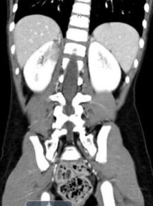
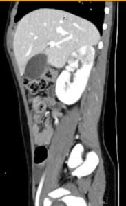
Large neuroblastoma mass above left kidney, distorting its architecture
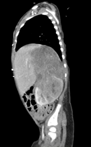
This image shows a large neuroblastoma mass at upper pole of left kidney, distorting its architecture
Right kidney upper pole defects on contrast, consistent with acute pyelonephritis
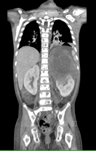
CT scan axial view with contrast showing right kidney upper pole defects, consistent with acute pyelonephritis
- « Previous
- 1
- 2