Radiology

Nuclear medicine imaging scan from back, showing a left pelvic kidney
May 11, 2021
KUB showing Prune Belly abdomen
May 11, 2021
Prenatal ultrasound showing distended bilateral renal hydronephrosis and distended urinary bladder (12 o’clock)
May 11, 2021
MRI image left kidney proximal ureter dilatation
May 11, 2021
CT scan showing bilateral renal cysts (arrows) in normal sized kidneys
May 11, 2021
CT scan coronal view with left ureter 2 mm stone (arrow)
May 11, 2021
MRI coronal view angiomyolipomas or hamartomas of tuberous sclerosis in right kidney
May 11, 2021
MRI image of left kidney lower pole angiomyolipoma of tuberous sclerosis
May 11, 2021
CTA showing renal artery stenosis
May 9, 2021

CTA showing right renal artery stenosis

Calyceal Diverticulum on MRU
April 20, 2021

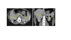
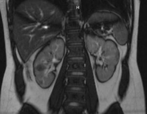
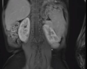
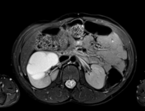
   Right anterior calyceal diverticulum on functional MR Urography. Top left side shows a T2 weighted image (no contrast) where fluid is bright, top right image is 10 min post-contrast on axial plane, and bottom images are a coronal views showing filling of diverticulum on dynamic phase. Images courtesy of…