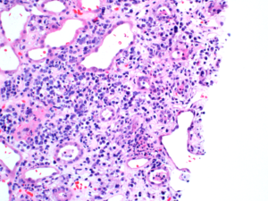
A mixed inflammatory cell infiltrate in a child with acute interstitial nephritis. Image courtesy of Patrick Walker, MD.
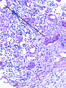
WBC casts (black arrows) in a biopsy specimen of a patient with acute interstitial nephritis. Images courtesy of Patrick Walker, MD.
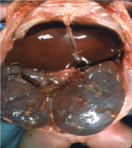
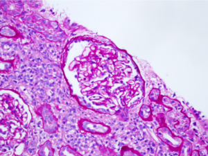
Gross pathology and low-power light microscopy of kidney tissue in a neonate with ARPKD. The kidneys are enlarged but maintain their reniform shape, and are full of microscopic cysts derived from dilated distal tubules and cortical collecting ducts. Images courtesy of Patrick Walker, MD.
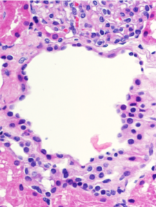
Microscopic renal cysts. Note the flattened to cuboidal epithelium lining the cysts. Images courtesy of Patrick Walker, MD.
Acute interstitial nephritis with associated acute tubular injury. There is interstitial edema and the tubules are not back to back as would be expected, due to the inflammatory and lymphocytic infiltrate in the interstitial compartment. Note the interstitial eosinophils present in the second image (black arrows). Images courtesy of Joseph…
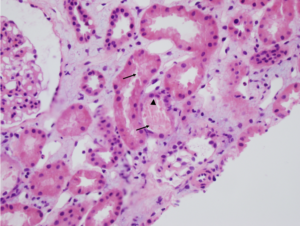
Acute tubular necrosis (ATN). Note the tubules are not back-to-back due to interstitial edema (Masson trichrome staining, not shown, did not show appreciable fibrosis). There is blebbing and sloughing of tubular epithelial cells (black arrows) with loss of the brush border, as well as flattening of the renal tubular epithelium…