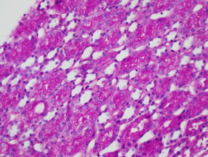
Numerous PAS-positive protein reabsorption droplets in the renal tubules of a child with minimal change disease. Image courtesy of Joseph Gaut, MD PhD.
IgA nephropathy with crescents. Note the fibrocellular crescent present in this glomerulus extending from the 3 o’clock the 12 o’clock position. Though difficult to appreciate in this image, there is mild mesangial hypercellularity. Image courtesy of Joseph Gaut, MD PhD.
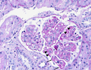
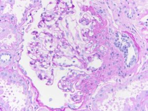
PAS stain of a glomerulus in a patient with SLE nephritis. There is endocapillary and mesangial proliferation, as evidenced by thickened, occlusive capillary loops and increased mesangial cellularity. Note the “wire loop” lesions (arrows) and hyaline thrombi present (arrowheads). Image courtesy of Joseph Gaut, MD, PhD.
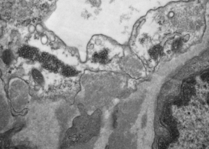
Tubuloreticular inclusions in a patient with diffuse proliferative SLE nephritis (SLE class IV). These subcellular structures (dark circular clusters) on transmission electron microscopy are localized to the cytoplasm of endothelial cells, and thought to be formed in high interferon states. These are classic for SLE nephritis, but can be seen…
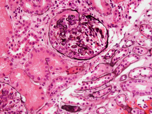
Segmental obliteration of the glomerular capillary lumen in a patient with FSGS. Note the sclerotic portion of the glomerular tuft is adherent to Bowman’s capsule. There is proximal tubular hypertrophy, which can be seen in this condition in response to heavy proteinuria. Image courtesy of Brian Stotter, MD.
- « Previous
- 1
- 2